FIGURE 1.

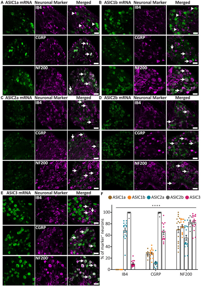
ASICs expression in naïve adult mouse DRG. (A–E) Representative confocal images showing the presence of ASIC1a (A), ASIC1b (B), ASIC2a (C), ASIC2b (D), and ASIC3 (E) transcripts in DRG neurons with different neuronal markers, including IB4, CGRP, and NF200. (F) Percentage of the marker positive neurons expressing different ASIC subunits. Arrow heads point to neurons expressing only ASIC1a, ASIC1b, or IB4. Arrows point to ASIC/marker-double positive neurons. The expression pattern of the five ASICs subunits was significantly different among the three neuronal subpopulations (****p < 0.0001, using two-way ANOVA). Scale bar = 50 μm; n = 9–19 DRGs from 3 to 4 naïve mice.
