
Figure 2.
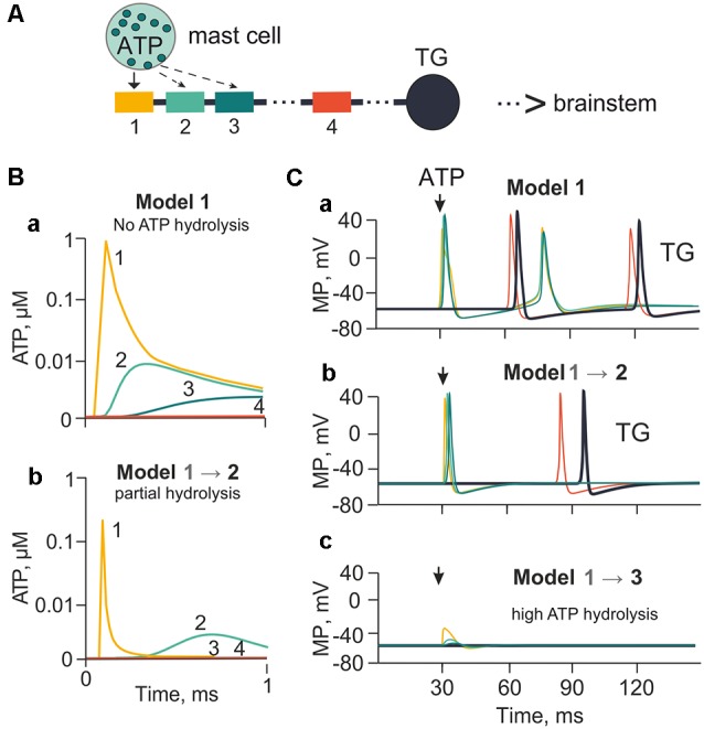
The model of meningeal afferents with different rates of ATP hydrolysis. (A) Schematic presentation of the multi-compartment model of neuro-immune synapse with ATP release from mast cells and acting to several neighboring nerve compartments. (Ba) ATP concentration via diffusion to each (1, 2, 3, 4) nerve compartment without hydrolysis. (Bb) ATP concentration at each compartment with partial hydrolysis (degradation coefficient 0.01). (Ca,b,c) Spiking activity with no ATP hydrolysis (Model 1), with low ATP hydrolysis (Model 2) and high rate of hydrolysis (degradation coefficient 0.8; Model 3), respectively.