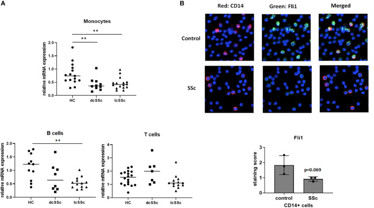
FIGURE 1.
Monocytes isolated from SSc patients have low levels of Fli1. (A) Monocytes, T and B lymphocytes were isolated from healthy controls (HC) and SSc patients (diffuse-dcSSc or limited-lcSSc) and mRNA levels of Fli1 were analyzed using quantitative RT-PCR. One way ANOVA (GraphPad Prims 7) was used to compare SSc to control. (B) Isolated peripheral blood mononuclear cells (PBMCs) from SSc and healthy controls (HC) were stained with CD14 for monocytes (red) and Fli1 (green) antibodies, and DAPI was used to counterstain the nuclei. Representative images and quantification are shown (n = 3 each). **p < 0.01.

