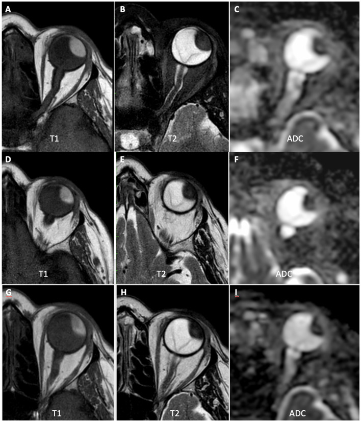
Figure 3.
A 66-year-old patient with choroidal melanoma in the left eye (upper temporal quadrant, measuring 15 × 14 × 9 mm), was treated with brachytherapy. (A–C) MR1 showing tumor with high signal at T1-weighted images, low signal at T2-weighted images, and restricted diffusion on the ADC map (mean ADC: 0.90 × 10⁻³ mm2/s). (D–F) MR2 showing that the tumor had similar morphological characteristics and dimensions, but with a slight increase in ADC values compared to the initial examination (mean ADC: 0.97 × 10⁻³ mm2/s). (G–I) MR3 showing a slight reduction in tumor dimensions, with further increase in ADC values (mean ADC: 1.20 × 10⁻³ mm2/s).

