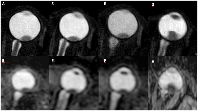
Figure 4.
A 59-year-old patient with choroidal melanoma in the left eye (upper temporal quadrant, measuring 9 × 4 mm), was submitted to brachytherapy. Pretreatment MR images: (A) axial T2-weighted sequence and (B) axial ADC map, with mean ADC of 1.241 × 10⁻³ mm2/s. MR images collected 3 months after treatment in the same sequences demonstrate stability of lesion size (C) and decreased ADC values (D) (mean ADC: 1.007 × 10⁻³mm2/s). MR images 6 months after treatment show stability of the lesion size (E) and ADC values (F). MR images collected 9 months after treatment show an increase in lesion dimensions (G) (13 × 9 mm) and decreased ADC values (H) (mean ADC: 0.794 × 10⁻³mm2/s).

