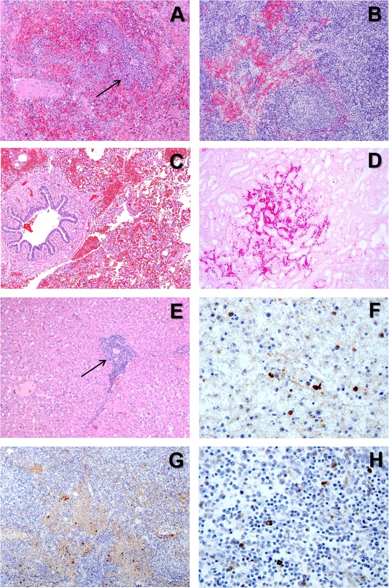
Figure 5.
(A) H&E stain of the spleen from a pig with acute ASF showing abundant red blood cells within the red pulp and severe lymphoid depletion, with very small lymphoid follicles (arrow) in the white pulp. (B) H&E stain of the gastrohepatic lymph node from a pig with subacute ASF showing hemorrhages in the perifollicular lymphoid tissue and the medulla, together with a moderate lymphoid depletion. (C) H&E stain of the lung from a pig with subacute ASF showing severe hemorrhages in the septa and the alveolar spaces. (D) H&E stain of the kidney from a pig with acute ASF showing interstitial hemorrhages within the renal cortex. (E) H&E stain of the liver from a pig with acute ASF showing periportal inflammatory infiltrates (arrow) composed of lymphocytes, macrophages and plasma cells. (F) IHC detection of ASFV p72 in the spleen showing strong positive reaction in macrophages in the red pulp and cell debris within the necrotic areas. (G) IHC detection of ASFV p72 in the gastrohepatic lymph node showing strong positive reaction in macrophages within the perifollicular areas and the medulla. (H) IHC detection of ASFV p72 in the tonsil showing strong positive reaction in macrophages within the perifollicular areas.

