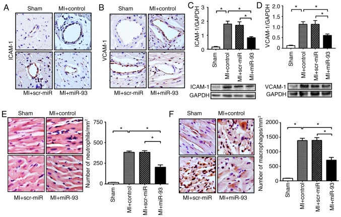
Figure 5.
Increased expression of miR-93 attenuates the infiltration of neutrophils and macrophages into the myocardium after MI. After 7 days of transfection, mice were subjected to MI for 3 days. Representative immunohistochemical images of ICAM-1 and VCAM-1 in the heart sections (magnification, ×400). Immunohistochemistry found a decrease in the positive staining of (A) ICAM-1 and (B) VCAM-1 in the ischemic myocardium, following transfected with lentivirus expressing miR-93 after MI. The dark brown color indicates positive staining. Western blot analysis showed the attenuation of (C) ICAM-1 and (D) VCAM-1 protein expression levels in miR-93 transfected hearts after MI. n=4-5 mice per group. Representative images of infiltrated neutrophils and macrophages in heart sections (magnification, ×400). Increased expression levels of miR-93 attenuated MI-induced infiltration of (E) neutrophils and (F) macrophages into the myocardium. The pink color indicates positive neutrophils. The dark brown color indicates positive macrophages. n=3 mice per group. Data are presented as the mean ± SEM. *P<0.05 vs. the indicated group. MI, myocardial infarction; scr, scrambled; miR, microRNA; ICAM-1, intercellular cell adhesion molecule-1; VCAM-1, vascular cell adhesion molecule-1.

