
Figure 1.
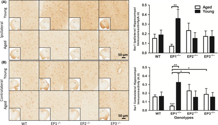
Effect of PGE2 EP1, EP2, and EP3 receptor deletion on microglial activation after rCHI. Seventy‐two hours after TBI, mice were sacrificed and brain sections were processed for Iba1 immunochemistry to evaluate hippocampal microgliosis and morphological changes. (A and B) Representative high magnification images of hippocampal brain sections showing the aged and young (A) ipsilateral and (B) contralateral hemispheres of WT, EP1−/−, EP2−/−, and EP3−/− mice. Square selections denote the magnified regions' locations. Quantification of brown positive pixel count indicated that EP1−/− aged mice had significantly less (A) ipsilateral and (B) contralateral Iba1 immunoreactivity compared with WT aged mice. (B) EP1−/− aged mice had significantly less Iba1 immunoreactivity compared with EP1−/− young mice. EP1−/− aged mice had significantly less Iba1 immunoreactivity compared with EP2−/− and EP3−/− aged mice. The reduction in microglial activation was accompanied by distinct morphological changes. Comparisons included aged male and young male wild type (n = 8, n = 7), EP1−/− (n = 10, n = 8), EP2−/− (n = 8, n = 9), EP3−/− (n = 8, n = 11), *P < .05, **P < .01