
Figure 2.
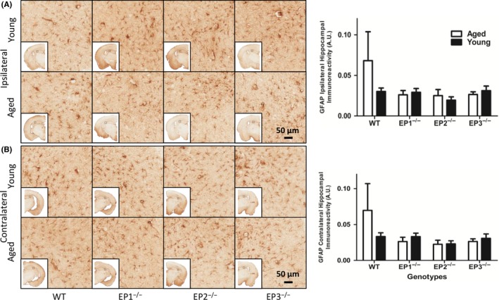
Effect of PGE2 EP1, EP2, and EP3 receptor deletion on astroglial activation after rCHI. Seventy‐two hours after TBI and WT, mice were sacrificed and brain sections were processed for GFAP immunochemistry to evaluate hippocampal astrogliosis and morphological changes. (A and B) Representative high magnification images of hippocampal brain sections showing the aged and young (A) ipsilateral and (B) contralateral for WT, EP1−/−, EP2−/−, and EP3−/− mice. Square selections denote the magnified regions' locations. Quantification of brown positive pixel count demonstrated that no significant difference in GFAP immunoreactivity was found in (A) ipsilateral and (B) contralateral regions across both genotype and age groups. Comparisons included aged male and young male wild type (n = 8, n = 7), EP1−/− (n = 10, n = 8), EP2−/− (n = 8, n = 9), and EP3−/− (n = 8, n = 11)