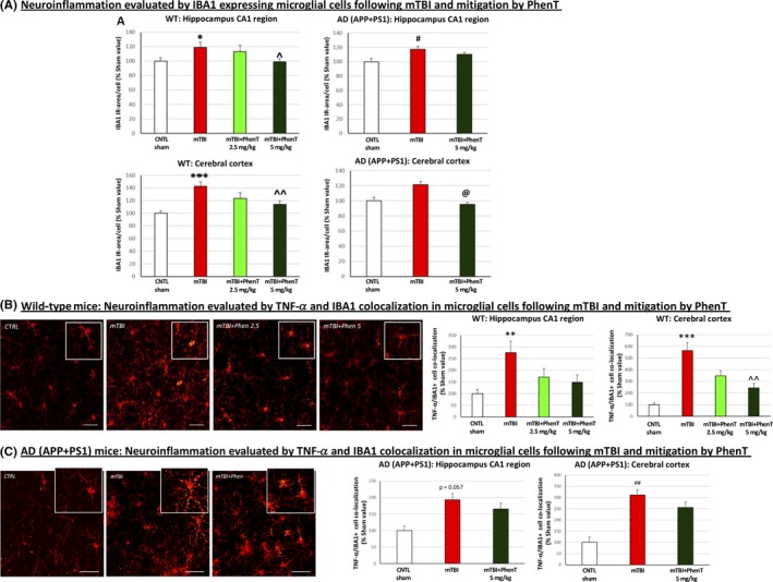
Figure 4.

PhenT mitigates markers of microglial neuroinflammation in mTBI‐challenged WT and AD mice across hippocampus and cerebral cortex (72 h post‐mTBI). After mTBI injury, evaluation of (A) IBA1 + cells (red in appearance) showed a rise in number in mTBI vehicle treated‐WT and AD vs control (CTRL) mice (w/o mTBI). PhenT mitigated this. A mTBI‐induced elevation in microglial cells co‐expressing TNF‐α (yellow in appearance) was also evident in (B) WT and (C) AD Tg mice, which was mitigated by PhenT treatment (n = 5/group). In the higher magnification insets in (B) and (C) IBA1 and TNF‐α are red and yellow, respectively (the TNF‐α is evident in small yellow punctate within the red colored IBA1 immunostained microglia). *P < .05, **P < .01, ****P < .0001 vs CTRL by Tukey's post hoc test; ^P < .05, ^^P < .01, ^^^P < .001, vs mTBI by Tukey's post hoc test. #P < .05 ##P < .01 vs CTRL by Mann‐Whitney rank test; @P < .05 vs mTBI by Mann‐Whitney rank test. Data shown as mean ± SEM Scale bar = 30 μm40
