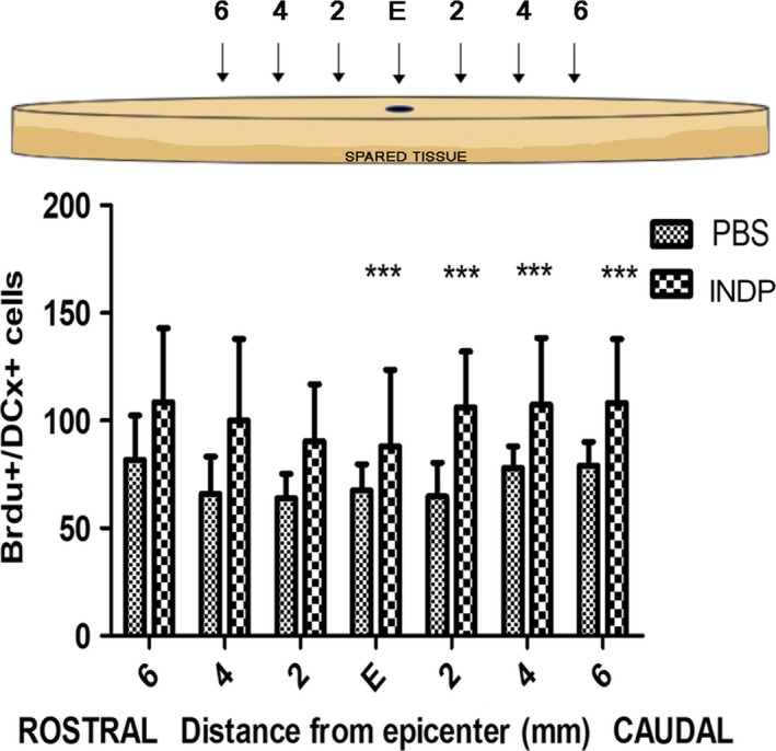
FIGURE 4.

Number of BrdU+/DCX+ cells at the epicenter (E), rostral, and caudal stumps of the SC. The INDP group showed a significant increase in the total number of BrdU/DCX+‐labeled cells (neuroblasts) compared to the PBS group. Bars represent the mean ± SD of five rats. This is one representative graph of three determinations. *Different from PBS, P < .05; one‐way ANOVA followed by Tukey's test
