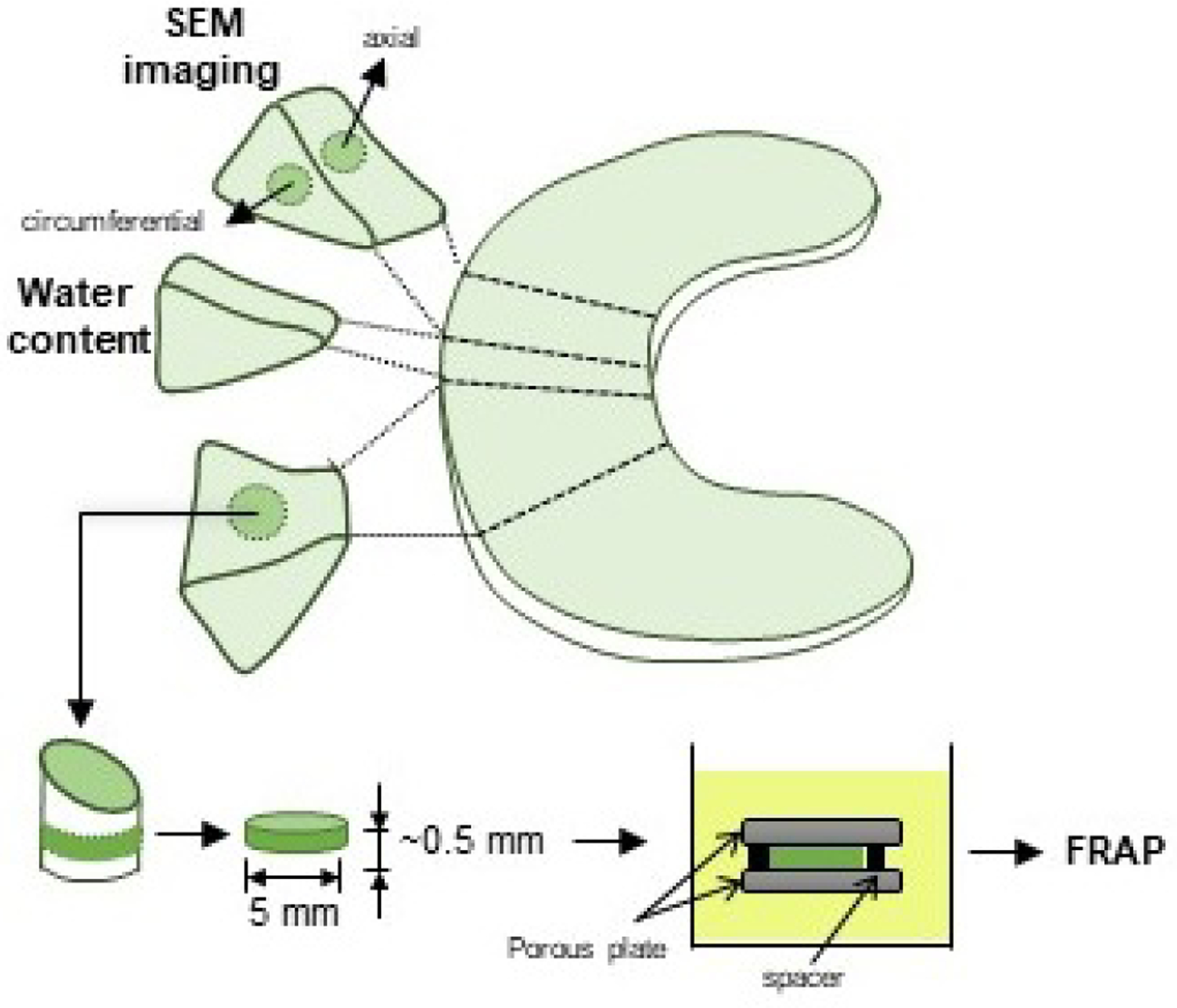
Figure 1:

Schematic of specimen preparation. Location and size of the specimens is shown. For FRAP tests, cylindrical specimens with a height of 0.5 mm and a diameter of 6 mm were prepared from the central region of the meniscus along the axial direction. For SEM imaging, specimens were prepared from central regions along the axial and the circumferential directions.
