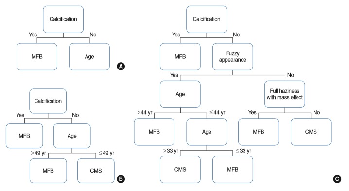
Fig. 3.
Regression tree model for diagnosis. (A) Model 1. (B) Model 2. (C) Model 3. Model 1 only involved intralesional hyperdensity as a variable; model 2 added demographic data; and model 3 included intralesional hyperdensity and demographic data, as well as radiological features (lobulated protruding lesion and full haziness with mass effect). Model 1: area under the curve, 0.853; accuracy, 81.78%; model 2: 0.880, 79.76%; model 3: 0.904, 88.66%, respectively. MFB, maxillary sinus fungus ball; CMS, chronic maxillary sinusitis.

