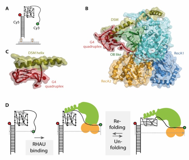
Figure 4.
(A) Schematic representation of one G-quadruplex (G4) single molecule Förster resonance energy transfer (smFRET) construct used in the described studies. (B) Co-crystal structure (pdb 5VHE) of RHAU in complex with a c-Myc G-quadruplex, color-coded for different important protein domains. (C) The DSM helix of RHAU stacks as planar, unipolar surface on top of the G-quadruplex. (D) Unfolding mechanism of DNA G4s by RHAU derived from smFRET studies. Binding of RHAU to the G4 results in FRET decrease, followed by ATP independent, repetitive partial unfolding and refolding of DNA G4. Figures A and D were adapted and modified from Tippana et al. and are used under Creative Commons Attribution 4.0 International License (CC-BY 4.0).

