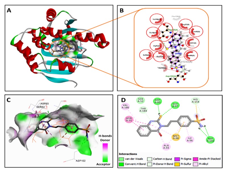
Figure 3.
2/3D binding modes of onalespib and HAA2020 into the active site of Hsp90 (pdb code: 2XJX): (A) binding modes of the redocked onalespib (coloured in yellow), co-crystallized onalespib (coloured in blue) and HAA2020 (coloured by element) into Hsp90, (B) LigPlot view showing the superposition of HAA2020 and onalespib with two equivalent hydrogen bonds (shown as olive green dotted lines) with Asp93 and Asp102 and several equivalent hydrophobic interactions (shown as brick red dotted lines), (C) 3D binding mode of HAA2020 (shown as sticks coloured by element) overlaid with onalespib (shown as orange lines) into the binding site of Hsp90, receptor shown as the hydrogen bond surface, hydrogen atoms omitted for clarity, and (D) 2D binding mode of HAA2020 showing three conventional hydrogen bonds and different types of hydrophobic interactions.

