Figure 4.
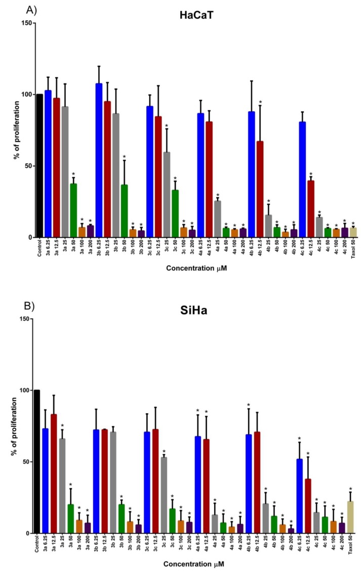
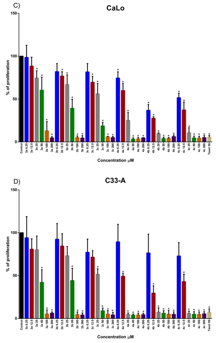
Proliferation effect of Naphthoquinone amino acid derivatives was evaluated in HPV positive cancer cell lines derived from cervix and a non-tumorigenic HPV negative cell line. (A) HaCaT HPV negative cell line cells were treated with 6.25, 12.5, 25, 50, 100 and 200 μM of naphthoquinone amino acid derivatives to assay proliferation rate at 72 h post-treatment. (B) SiHa HPV 16 positive cancer cells were treated with 6.25, 12.5, 25, 50, 100 and 200 μM of naphthoquinone amino acid derivatives to assay proliferation rate at 72 h post-treatment. (C) CaLo HPV 18 positive cancer cells were treated with 6.25, 12.5, 25, 50, 100 and 200 μM of naphthoquinone amino acid derivatives to assay proliferation rate at 72 h post-treatment. (D) C33-A HPV negative cancer cells were treated with 6.25, 12.5, 25, 50, 100 and 200 μM of naphthoquinone amino acid derivatives to assay proliferation rate at 72 h post-treatment. Cells treated with 0.1% of DMSO were used as Control. * represents statistically significant p < 0.05 value.


