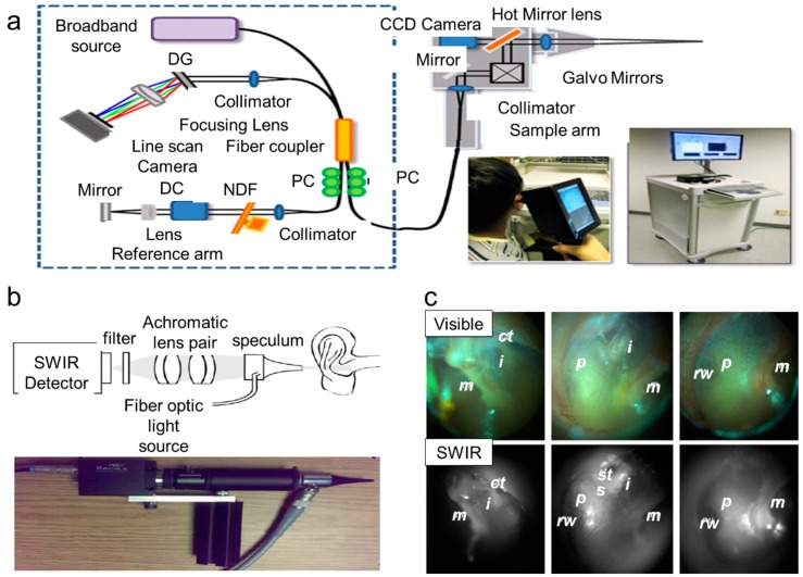
Figure 5.
Tools available to detect middle ear infection. (a) Handheld OCT (optical coherence tomography) (diffraction grating (DG); polarization controller (PC); dispersion compensation (DC) materials; neutral density filter (NDF)). Reproduced with permission from [76]. Copyright Elsevier B.V.,2013. (b) SWIR (short wavelength infrared) otoscope. Reproduced with permission from [81]. Copyright National Academy of Sciences,2016 (c) Representative images for (b) Reproduced with permission from [81]. Copyright National Academy of Sciences,2016. ct, chorda tympani; i, incus; m, malleus; p, cochlear promontory; st, stapedial tendon; s, stapes; rw, round window niche.

