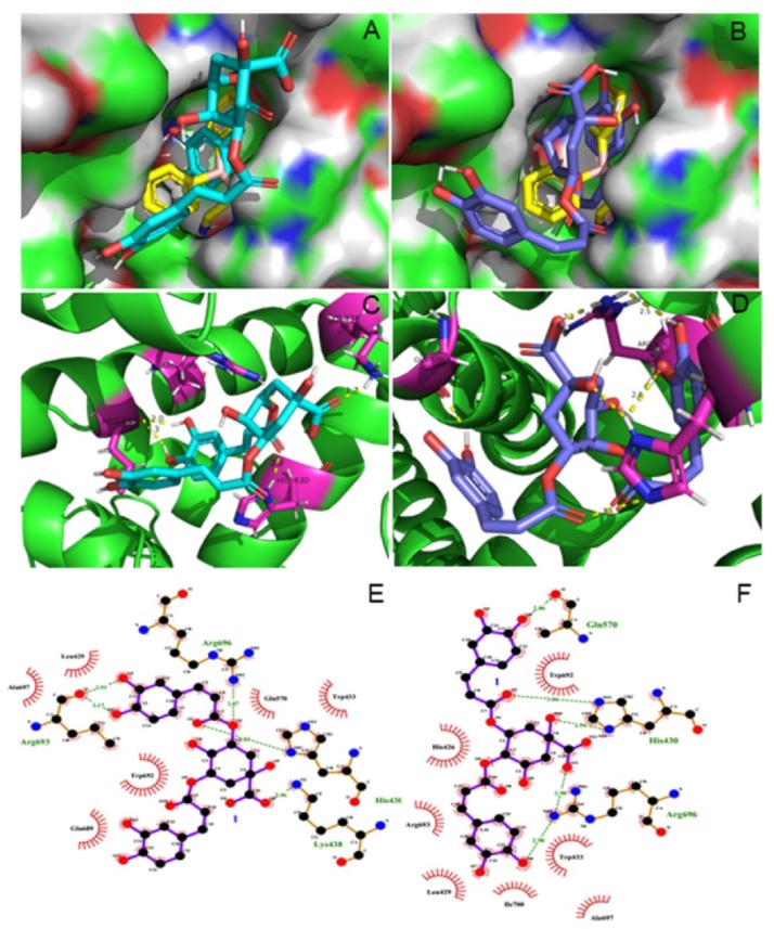
Figure 3.

Docked conformations of isochlorogenic acid (A), isochlorogenic acid (B), and agonist 2-APB with cyan, slate, and yellow sticks into the active site of 6MHO protein, respectively (A,B). Analysis of molecular docking showing the key interactions in the binding pocket. The key residues are presented in the form of purple sticks, and hydrogen bonds are drawn with dotted yellow (C,D). Ligplot+ of isochlorogenic acids A and B bound to 6MHO protein showing the key hydrophobic interactions (E,F).
