
Figure 7.
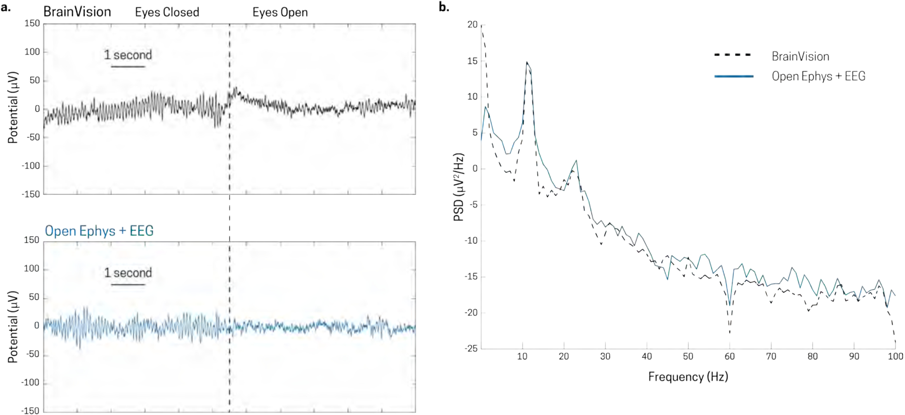
Signal comparison between Brainvision actiCHamp system and Open Ephys + EEG system using the same Brainvision actiCap electrode cap in recording eyes closed alpha activity. Transition from eyes closed to eyes open in the [top figure] Brainvision system and the [bottom figure] Open Ephys + EEG are very similar. b) Average PSD of Brainvision (dotted black line) and Open Ephys + EEG (solid blue line) for eyes closed epochs.