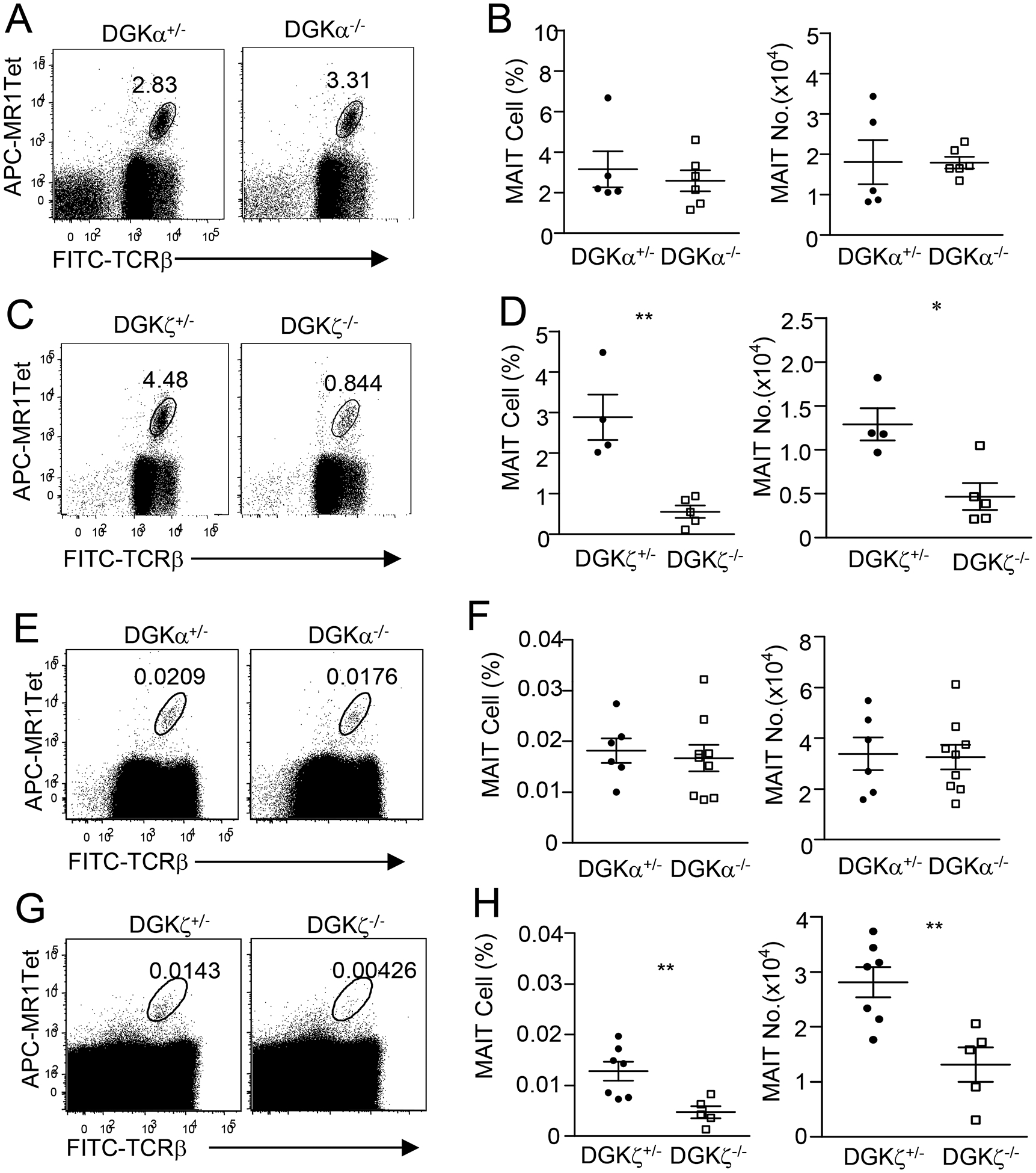
Figure 5. Effects of DGKα or ζ deficiency on thymic MAIT cell numbers.

Thymocytes from 8–10-week-old mice enriched for MAIT cells with MR1-Tet (A–D) or total thymocytes (E–H) were stained with fluorescently labeled anti-TCRβ, MR1-Tet, and other antibodies similar to Figure 1. A, C, E, H. Dot plots show TCRβ and MR1-Tet straining in live gated Lin− cells from Dgka−/− and Dgka+/− control mice (A,E) or Dgkz−/− and Dgkz+/− control mice (C, G). B, D, F, H. Scatter plots represent mean ± SEM of MAIT cell percentages and numbers of Dgka−/− (B,F) and Dgkz−/− (D,H) mice and control mice. For A and B, data shown are representative of or pooled from five experiments (N = 5 for DGKα+/−, N = 6 for DGKα−/−). For C and D, data shown are representative of or pooled from four experiments (N = 4 for DGKζ+/−, N = 5 for DGKζ−/−). For E and F, data shown are representative of or pooled from six experiments (N = 6 for DGKα+/−, N = 9 for DGKα−/−). For G and H, data shown are representative of or pooled from five experiments (N = 7 for DGKζ+/−, N = 5 for DGKζ−/−). Dgka+/− and Dgkz+/− mice had similar numbers of MAIT cells to WT mice and were used as control. *, p<0.05; **, p<0.01 determined by unpaired Student t-test.
