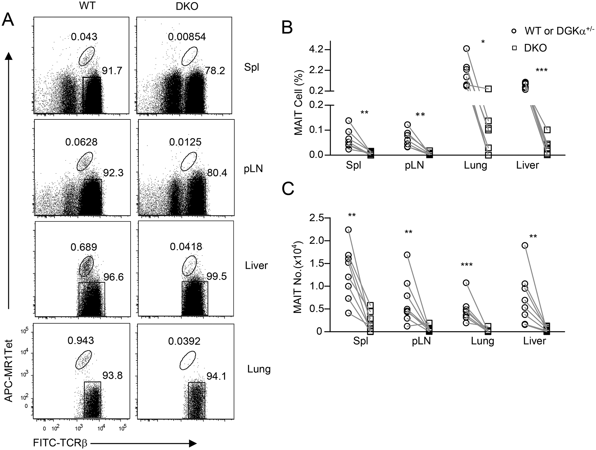
Figure 7. Severe decreases of MAIT cells in the peripheral organs in DGKαζ DKO mice.

Single cell suspensions of the spleen, pLNs, lung, and MNCs of the liver from 8–10-week-old Dgka−/−Dgkzf/f-CD4Cre (DKO) and DGKα+/− or WT control mice were directly stained with MR1-Tet, anti-TCRβ, CD44, CD24, CD45, and lineage antibodies. A. Dot plots show TCRβ and MR1-Tet straining in live gated Lin− cells. For lung and liver, only TCRβ+ cells were gated and shown. B. MAIT cell percentages. C. MAIT cell numbers. Data shown are representative or pooled from eight experiments (N = 8 for both control and DKO mice). Each circle and square represents one control and DKO mouse respectively. *, P<0.05; **, P<0.01; ***, P<0.001 determined by pairwise Student t-test.
