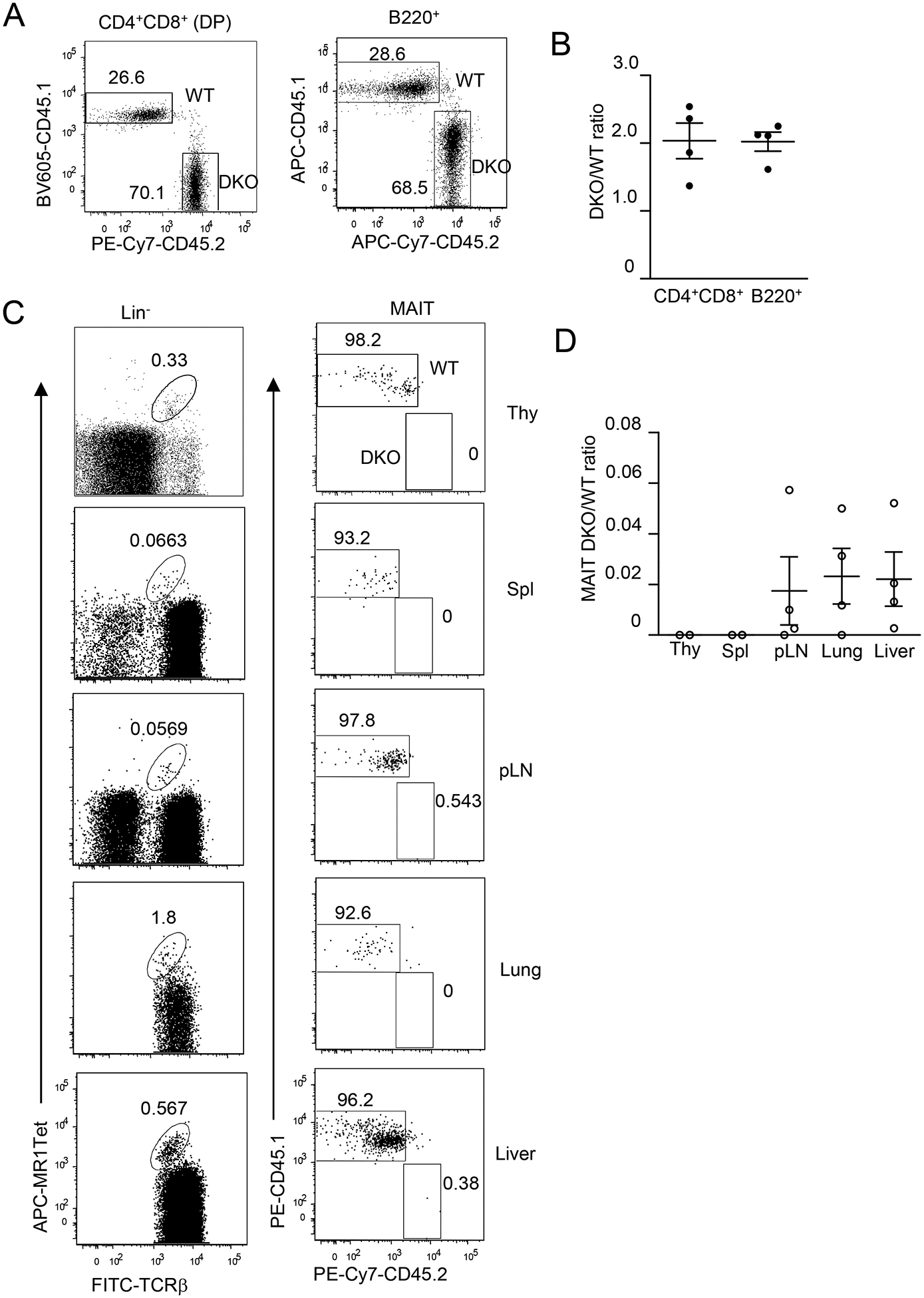
Figure 8. DGKα and ζ intrinsically promote MAIT cell development.

Irradiated CD45.1+CD45.2+ WT recipient mice were reconstituted with CD45.1+ WT and CD45.2+ DKO BM cells and were analyzed 8 weeks later. A. Representative dot plots showing CD45.1 and CD45.2 staining in live gated CD4+CD8+ DP thymocytes and B220+ splenocytes. B. DKO to WT ratios of DP thymocytes and B220+ splenic B cells. C. Representative dot plots showing MAIT cell staining in live gated Lin− cells (Thy, Spl, and pLNs) or in Lin−TCRβ+ cells (lung and liver) and CD45.1 and CD45.2 staining in MAIT cells. D. DKO to WT ratios of MAIT cells. Data shown represent or are pooled from four experiments with one recipient mouse examined in each experiment. Each circle represents one recipient mouse. Horizontal bars represent mean ± SEM (N = 4).
