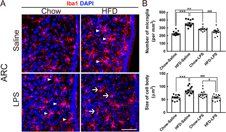
Fig. 5.
Morphological alteration of microglia in the hypothalamic arcuate nucleus was induced by intermittent LPS administration into HFD-fed mice. a Brain tissue sections containing the hypothalamic arcuate nucleus (ARC) were prepared from animals from the four groups after feeding for 5 months and then subjected to Iba1 immunofluorescence (red) and DAPI nuclear counterstaining (blue). Representative active microglia with hypertrophic shapes are indicated by arrowheads. Arrows indicate microglia with a small size and fine processes in the HFD-LPS group. b The number of Iba1+ microglia accumulated in the ARC (per mm2) in the four groups was quantified. In addition, the average cell body size of ARC microglia was measured. The data are presented as the mean ± SEM (n = 12 tissue sections from 3 animals from each group). **p < 0.01, ***p < 0.001 versus the chow-saline group; #p < 0.05, ###p < 0.001 versus the HFD-LPS group. Scale bar in A = 50 μm

