Fig. 1.
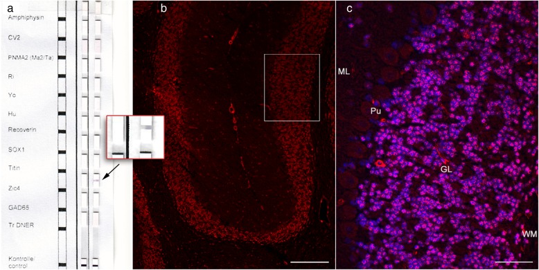
Immunoblot and Immunofluorescence Test on Mouse Cerebellum. Demonstration of Zic4 antibodies in serum (the same results were obtained with cerebrospinal fluid, not shown). a On an immunoblot, 1:100 diluted patient serum produces on the right lane a band with Zic4 only (arrow and insert), not with any other antigen (Euroline DL 1111–1601-4 G, Euroimmun, Lübeck, Germany). The left lane was incubated with a control serum. b Incubation of patient serum, diluted 1:20, with unfixed mouse cerebellum (Euroimmun, Lübeck, Germany). Patient’s immunoglobulin G (IgG) binds in a Zic-4-typical pattern to the cerebellar granular cells (cf. Fig. 1 in Bataller et al., 2002 [5]). Bound antibodies are visualized by an anti-human-IgG antibody coupled to a red fluorochrome. Original magnification × 100. Bar: 100 μm. The area in the rectangle is shown in (C). c Original magnification × 400. Bar: 25 μm. Zic-4 IgG-antibodies bind to the nuclei of the granular layer and spare the nucleoli. Nuclear counterstaining with Hoechst 33342 1:10.000 in blue. ML = molecular layer; Pu = Purkinje cell layer; GL = granular layer; WM = white matter

