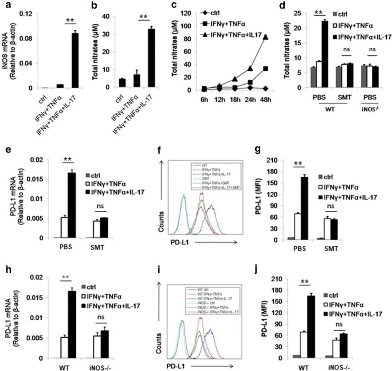
Fig. 2.
IL-17 enhances the expression of PD-L1 through NO. a C57BL/6 MSCs were treated by indicated cytokines for 24 h and then collected for the measurement of NO mRNA by real-time PCR. b, c C57BL/6 MSCs were treated as in A and after 24 h (b) or indicated time points (c), supernatant were collected for the assay of NO concentration by Griess reagent. d WT MSCs (C57BL/6 MSCs) and iNOS−/− MSCs were treated by cytokines and SMT (20 mM) for 24 h and supernatant were collected for the measurement of NO. e, f C57BL/6 MSCs were treated by cytokines with or without SMT. After 24 h, cells were collected for the assay of PD-L1 mRNA by real-time PCR. After 48 h, cells were collected for the assay of PD-L1 protein by flow cytometry. g The statistical results of three separate experiments described in f are also showed. h, i WT MSCs and iNOS−/− MSCs were treated with cytokines. Cells were collected for the measurement of PD-L1 mRNA and protein after 24 h and 48 h, respectively. j The statistical results of three separate experiments described in i are also showed

