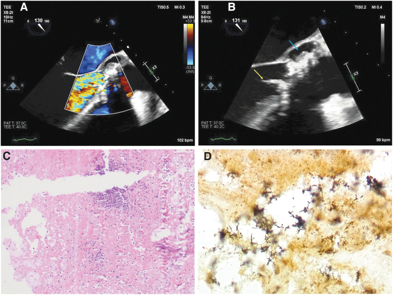
FIG. 1.
Aortic valve echocardiography and surgical pathology. (A) Transesophageal echocardiogram midesophageal atrioventricular long axis view with Doppler of the aortic valve demonstrating severe regurgitation (yellow-orange-red turbulent flow). (B) Transesophageal echocardiogram midesophageal long axis view of the calcified bicuspid aortic valve with mobile vegetation (yellow arrow) and perivalvular abscess (blue arrow). (C) H&E stain at 10 × magnification demonstrating chronic inflammation. (D) Steiner stain at 100 × magnification demonstrating abundant pleomorphic rods. H&E, hematoxylin and eosin. Color images are available online.

