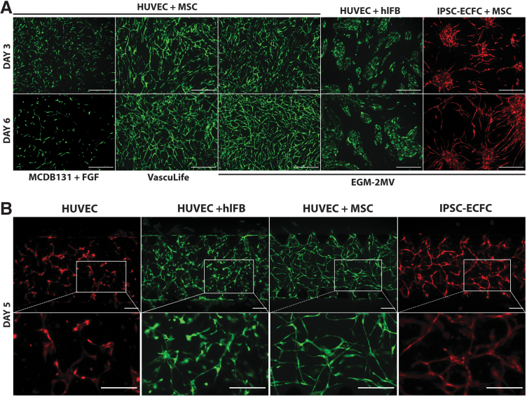
FIG. 1.
Effect of cell types and culture medium on microvascular network formation in 2D and 3D. (A) Representative images of vascular networks formed by HUVECs and iPSC-ECFC cocultured with MSCs or human lung fibroblasts in different culture media. Images were taken after 3 and 6 days of culture. Scale bars, 500 μm. (B) Representative images of vascular networks formed by HUVECs alone, HUVECs+human lung fibroblasts, HUVECs+adipose-derived MSCs, and iPSC-ECFC+adipose-derived MSCs in a fibrin gel. Images were taken after 5 days of culture. Scale bars, 200 μm. 2D, two-dimensional; 3D, three-dimensional; HUVECs, human umbilical vein endothelial cells; iPSC-ECFC, induced pluripotent stem cell-derived endothelial colony-forming cell; MSCs, mesenchymal stem cells.

