
Fig. 3 |. Lipid antigen presentation by thymocytes and DCs from ASM-deficient mice is reduced and bone marrow transfer restores iNKT cell levels in Asm−/− mice.
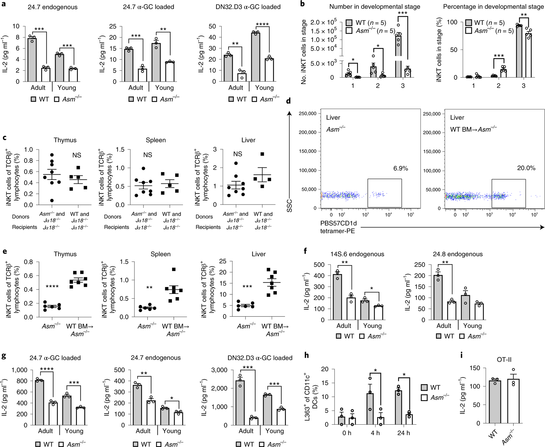
a, Thymocytes were incubated with α-GalCer (α-GC) for 4 h or left untreated, followed by addition of the indicated iNKT hybridomas. Thymocytes from both young (2-week-old) and adult Asm−/− mice were used as indicated. b, The graphs show absolute (left) and relative (right) numbers of iNKT cells in different thymic developmental stages in Asm−/− and WT mice. Stage 1 was defined as CD24loCD44loNK1.1lo, stage 2 as CD24loCD44hiNK1.1lo and stage 3 as CD24loCD44hiNK1.1hi. The results are representative of two independent experiments. c, Bone marrow (BM) chimeras were made by the transfer of a mixture of Jα18−/− and WT (n = 5) or Asm−/− (n = 8) BM into irradiated Jα18−/− recipients. The graphs demonstrate the percentage of cells positive for PBS57-loaded CD1d tetramer among TCRβ-positive cells (iNKT cells) 3 months after BM transfer. The results are representative of two independent experiments. d,e, Bone marrow (BM) chimeras were made by transfer of WT CD45.1+ BM to Asm−/− mice (n = 7) in comparison to non-irradiated Asm−/− mice not receiving BM (n = 6). The dot plots (d) show representative plots from the livers of a non-irradiated Asm−/− mouse not receiving BM and an Asm−/− mouse receiving WT BM. The graphs (e) demonstrate the percentage of cells positive for PBS57-loaded CD1d tetramer among TCRβ-positive cells (iNKT cells) 3 months after BM transfer. The results represent pooled results from three independent experiments. f,g, CD11c+ DCs were extracted from spleens with magnetic beads and co-cultured with the indicated NKT hybridomas. Before co-culture, the DCs in 24.7 α-GC loaded and DN32.D3 α-GC loaded were loaded with α-GC for 4 h (g). DCs from both young (2-week-old) and adult Asm−/− mice were used as indicated. h, CD11c+ DCs were extracted from spleens and incubated with α-GC for 4 or 24 h and stained with an antibody recognizing α-GC bound to CD1d in three technical replicates. The results are representative of two independent experiments. i, DCs from WT and Asm−/− mice were loaded with ovalbumin and cultured with ovalbumin-reactive T cells from OT-II mice. In all co-culture experiments (a,f,g and i), IL-2 levels were measured in three independent wells 20–24 h after addition of the indicated NKT hybridomas or T cells, and all these results are representative of three independent experiments. In all panels, the mean values are shown with the error bars representing the s.e.m. P values were calculated by a two-sided Student’s t-test in all panels except h, where a two-way ANOVA with Bonferroni’s correction for multiple comparisons was used. *P < 0.05, **P < 0.01, ***P < 0.001, ****P < 0.0001; NS, not significant.