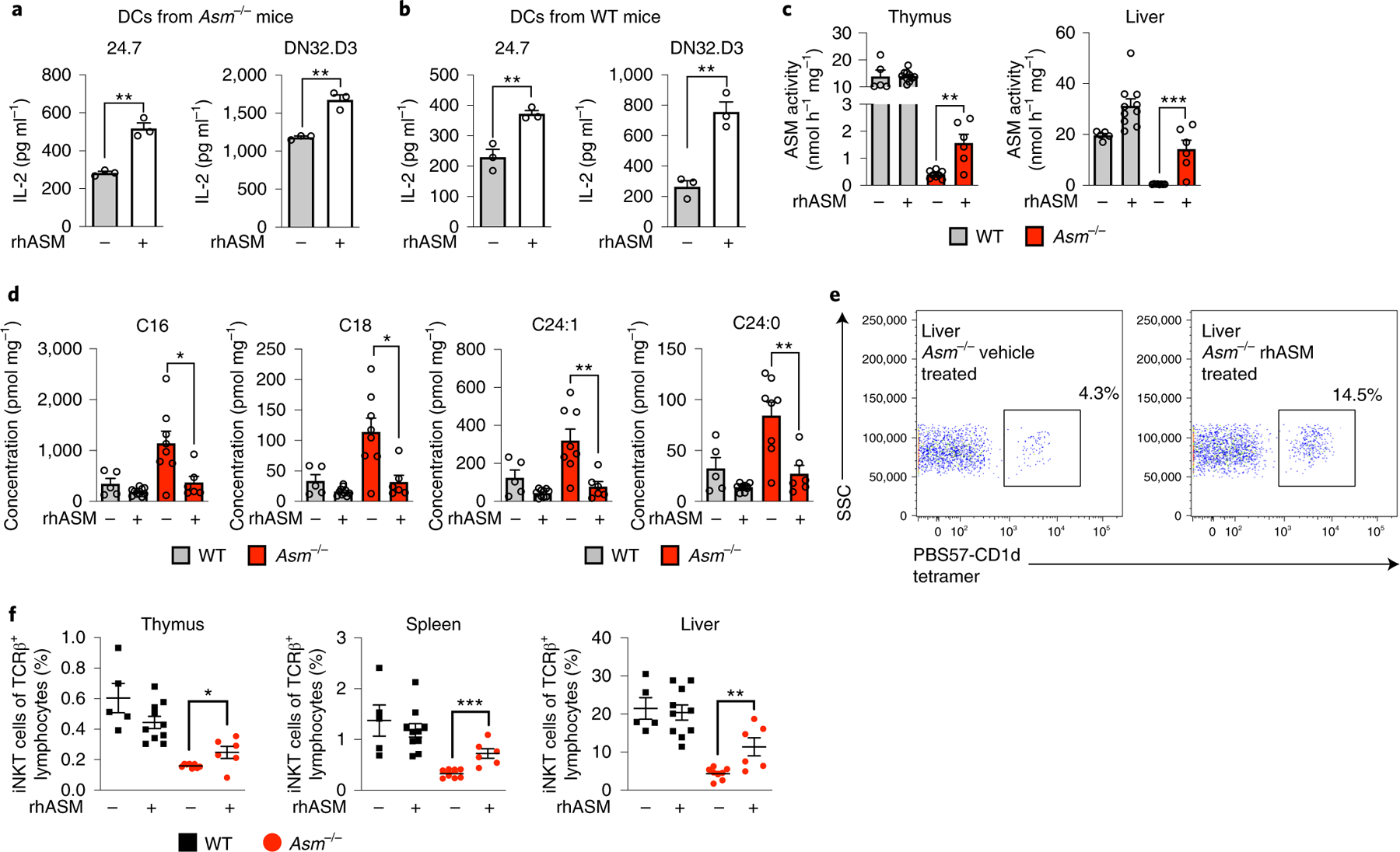
Fig. 8 |. Pharmacological ASM treatment in Asm−/− mice restores iNKT cells.

a,b, CD11c+ DCs were extracted from the spleens of Asm−/− mice (a) or WT (b) mice treated with one dose of rhASM (5 μg g−1) 12–16 h before extraction and loaded with α-GalCer for 4 h. The indicated iNKT hybridomas were added and IL-2 levels were measured in three independent wells after 20–24 h. The results are representative of three independent experiments. c, WT and Asm−/− mice were treated every other day from birth with rhASM (5 μg g−1). The graphs show the ASM activity levels in the liver and thymus of vehicle-treated and rhASM-treated Asm−/− and WT mice 2 d after the last enzyme injection. d, The graphs show the levels of the blocking sphingomyelin species as measured by mass spectrometry after treatment with rhASM. e, Representative dot plot from the livers of a vehicle-treated and an rhASM-treated Asm−/− mouse. The cells are gated on the lymphocyte population and TCRβ-positive cells. The percentages correspond to cells positive for PBS57-loaded CD1d tetramer. f, iNKT cell levels at the age of 2 weeks in the thymus, spleen and liver of mice that were treated with rhASM or vehicle as indicated in the graphs. The numbers represent the pooled results from three independent experiments with vehicle-treated (Asm−/− (n = 8) and WT (n = 5)) and rhASM-treated (Asm−/− (n = 6) and WT (n = 10)) mice (c,d and f). The hypothesis tested using the Student’s t-test was that rhASM would increase iNKT cell abundance in Asm−/− mice. In all panels, the mean values are shown with the error bars representing the s.e.m. P values were calculated by a two-sided Student’s t-test. *P < 0.05, **P < 0.01, ***P < 0.001.
