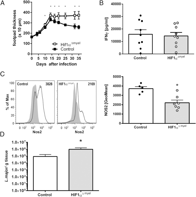
FIGURE 5.
HIF-1α stabilization in myeloid cells contributes to antileishmanial control in vivo. (A–D) Hif1αΔmyel mice and littermates (controls) were infected with L. major in their hind footpads. (A) Clinical course of cutaneous L. major infection (mean ± 95% confidence interval, n ≥ 3, representative of three similar independent experiments). *p < 0.01, Student t test or Mann–Whitney U test. (B) At day 28–29 postinfection, restimulation of draining lymph node cells from L. major–infected mice with soluble Leishmania Ag was performed. IFN-γ was determined in the culture supernatants (mean + SEM, n = 9 biological samples from two independent experiments). Student t test was performed and showed no significant difference. (C) At day 29 postinfection, NOS2 protein expression was determined in lesional CD11b+ cells from Hif1αΔmyel mice and littermates (control). Representative line graphs of NOS2 expression in lesional CD11b+ cells (left panels). Black line: NOS2 expression. Shaded area: isotype control. Geometric mean fluorescence of NOS2 (mean + SEM, n = 5–7) (right panel). A representative of two independent experiments is shown. *p < 0.05, Student t test. (D) At day 32 postinfection, L. major burden in skin lesion of infected mice was analyzed (mean + SEM, n = 8). A representative of two similar independent experiments is shown. *p < 0.05 Mann–Whitney U test.

