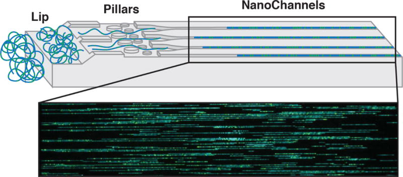
Fig. 2. Chip nanochannel structure and DNA loading for next-generation genome mapping (NGM).

The labeled double-stranded DNA is loaded into two flow cells (Irys or Saphyr, Bionano Genomics). The applied voltage concentrates the coiled DNA at the lip (left). Later, DNA is pushed through pillars (middle) to uncoil/straighten, then into nanochannels (right). DNA is stopped and imaged in the nanochannels. Blue - staining of DNA backbone. Green - fluorescently labeled nicked sites.
Figure originally published in Barseghyan et al. 2017, Genome Medicine 9:90. DOI 10.1186/s13073-017-0479-0. Reproduced here courtesy of publisher BioMed Central’s policy of free sharing of its open access articles.
