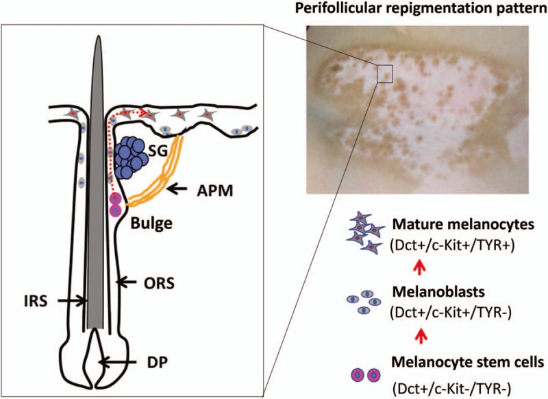
Figure 1.

Schematic depiction of cellular events underlying the perifollicular re-pigmentation pattern. (Upper-right) A male patient with vitiligo on the right side of his neck was treated twice a week with 308 nm excimer light (EL) for 2 months, after which multiple pigmented dots were seen around the hair follicles. (Left) Schematic of the bulge region in hair follicles showing a relatively safe niche that houses melanocyte stem cells (Dct+/c-Kit−/TYR−). Those melanocyte stem cells are mobilized to exit the bulge region upon exposure to narrow-band UVB (NB-UVB) or/308 nm EL and to start their differentiation or proliferation program to become melanoblasts (Dct+/c-Kit+/TYR−, also called transit amplifying [TA] cells). These melanoblasts continuously migrate upward through the ORS to the lesional epidermis and eventually differentiate into functional melanocytes (Dct+/c-Kit+/TYR+). The red dotted line indicates the putative migration route of melanocyte stem cells. DP: Dermal papilla; IRS: Inner root sheath; ORS: Outer root sheath; APM: Arrector pili muscle; SG: Sebaceous gland.
