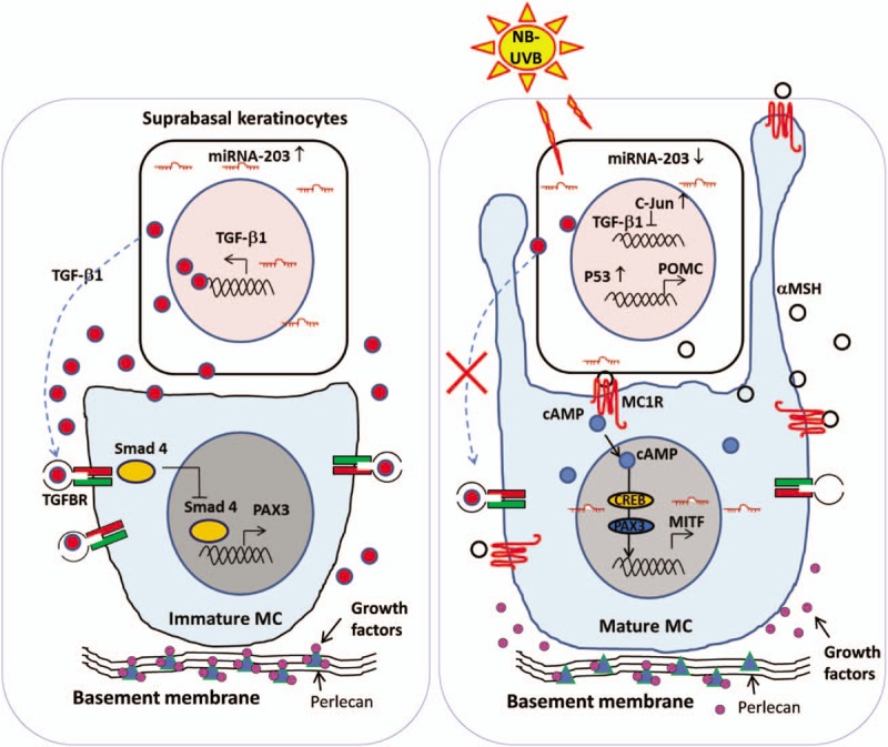
Figure 3.

The micro-environment favorable for re-pigmentation. (Left panel) In the absence of NB-UVB irradiation, differentiated keratinocytes residing in the suprabasal layers of the skin express high levels of miR-203 via a transcriptional activation mechanism. miR-203 post-transcriptionally represses c-Jun and then increases TGF-β1 expression, forming a TGF-β1-enriched extracellular micro-environment. (Right panel) In response to NB-UVB, keratinocytes activate the classical pathway of the p53/αMSH/MC1R/MITF cascade and suppress TGF-β1-ligand secretion via the miR-203/c-Jun pathway, which stimulates the proliferation and differentiation of melanocytes. NB-UVB also activates heparanase to induce the release of heparan sulfate-binding growth factors, which synergistically facilitates the proliferation and melanogenesis of melanocytes. All references are cited in the text. α-MSH: α-Melanocyte-stimulating hormone; cAMP: Cyclic adenosine monophosphate; MC: Melanocytes; MITF: Microphthalmia-associated transcription factor; NB-UVB: Narrow band ultraviolet B; POMC: Proopiomelanocortin; TGF-β1: Transforming growth factor beta-1.
