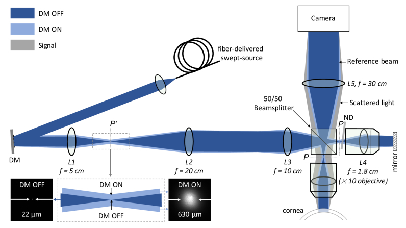
Fig. 1.
Fourier-domain full-field OCT system for in vivo corneal imaging of the human eye. DM – deformable membrane; ND – neutral density filter; P’ – plane conjugate to the pupil plane, P of the objective lens, L4; Dark blue shows the spatially coherent beam (when DM is OFF) and light blue – the spatially incoherent (when DM is ON). Gray beam indicates signal coming from the cornea that is backscattered at a range of angles. The beam is focused to a spot of 22 µm in the P’ plane in the coherent case (when DM is OFF), which is broadened to 630 µm in the incoherent case (when DM is ON), as shown zoomed-in in the inset.

