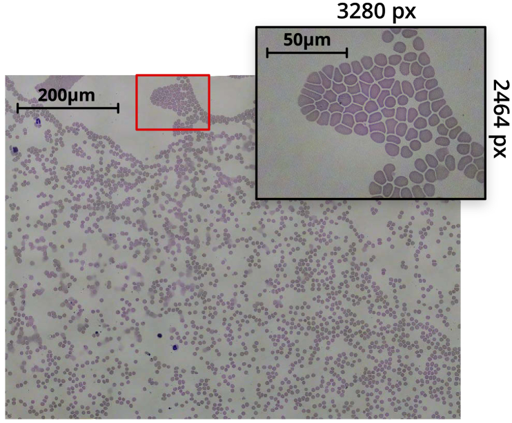
Fig. 4.
Tiled scan image of a Giemsa-stained thin blood smear, obtained with a , NA oil immersion objective. The inset highlights an individual 8-megapixel image from the scan. The composite image was obtained from a grid of captures. After accounting for image overlap and skewing, and cropping out edges of the composite with missing sections, the resulting image is px px ( megapixel).

