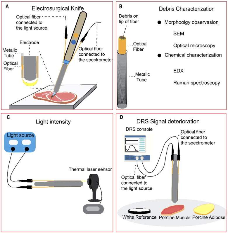
Fig. 1.
A) The design of the electrosurgical knife integrated with DRS (fiber-fiber distance = 3 mm). B) Characterizing the debris formed on the tip of optical fibers due to electrosurgery. C) The used setup to measure the intensity of light passing through the clean and used optical fibers. D) The setup to investigate the DRS signal deterioration after electrosurgery with three different sets of optical fiber, Clean Fiber (unused fiber), Muscle Fiber (contaminated with muscle tissue), and Fat Fiber (contaminated with adipose tissue) on three different surfaces (white reference, porcine muscle, and porcine adipose tissue).

