
Fig. 2.
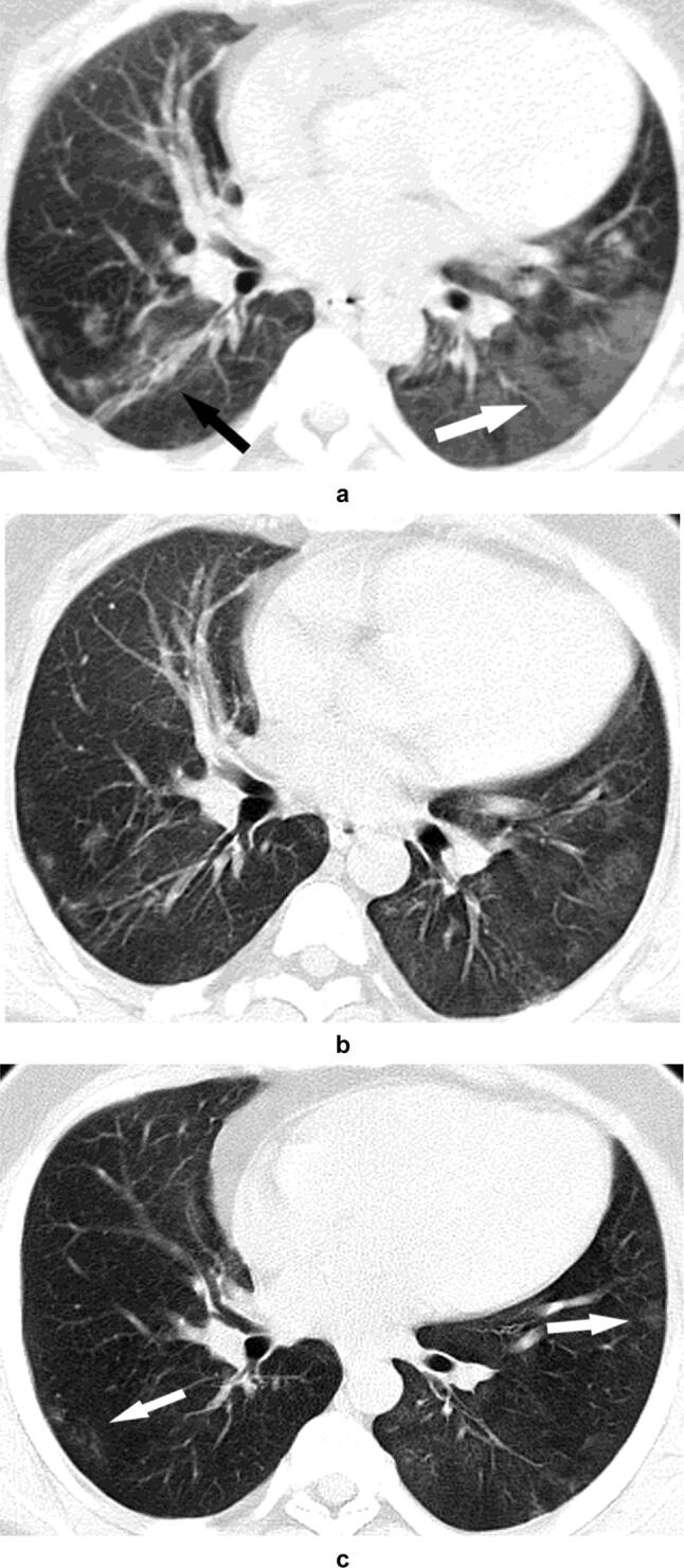
Patient 1. Images in a 28-year-old male with fever and fatigue. a On the admission day, the unenhanced CT scan shows diffuse bilateral multiple patchy GGO (white arrow), and the partial boundary is clear while some have unclear boundaries, which are especially significant in the lower lobes of both lungs; strip consolidative opacities (black arrow) are in the focal area. b On day 5, the follow-up unenhanced CT scan shows a significant reduction in GGO in both lungs, and solid shadows have no new lesions. c On day 11, the follow-up unenhanced CT scan shows that the lesion was basically absorbed, and few GGOs remain (white arrows). On day 15, the patient was discharged from the hospital