
Fig. 3.
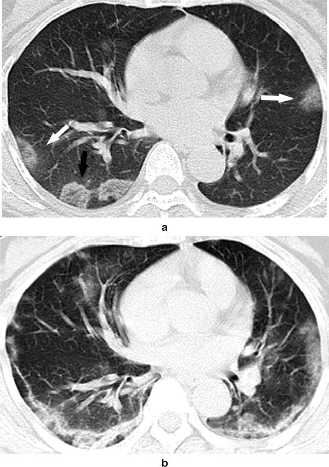
Patient 4. Images in a 57-year-old woman with fever. The DR is normal in the lung on the admission day. a The unenhanced CT shows multiple patchy GGO-like lesions in the both lungs at subpleural position, unclear boundaries of GGO shadows with halo signs (white arrows) or clear boundaries of partial glass density shadows with reversed halo signs (black arrows). The mesh shadow is visible inside (black arrow). b Follow-up unenhanced CT on day 12 shows multiple new lesions. The lesions spread and fused in the subpleural area, showing a crescentic shape, while some lesions changed in consolidation and partial fibrosis