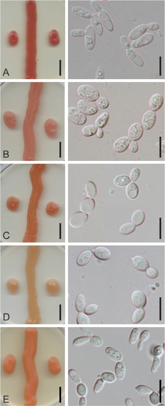
Fig. 2.

Colony and cell morphology of Symmetrospora species on YMA (left panels) and YM broth (right panels): A. Symmetrospora clarorosea strain SA308. B. Symmetrospora oryzicola strain MCA 4496. C–D. Symmetrospora pseudomarina strains SA42 (C) and SA716 (D, ex-type), showing the variation in colony color between the two strains. E. Symmetrospora suhii strain BG 02-5-27-3-2-2 (ex-type). Scale bars = 1 cm in culture images (left panels), 10 μm in cell images (right panels).
