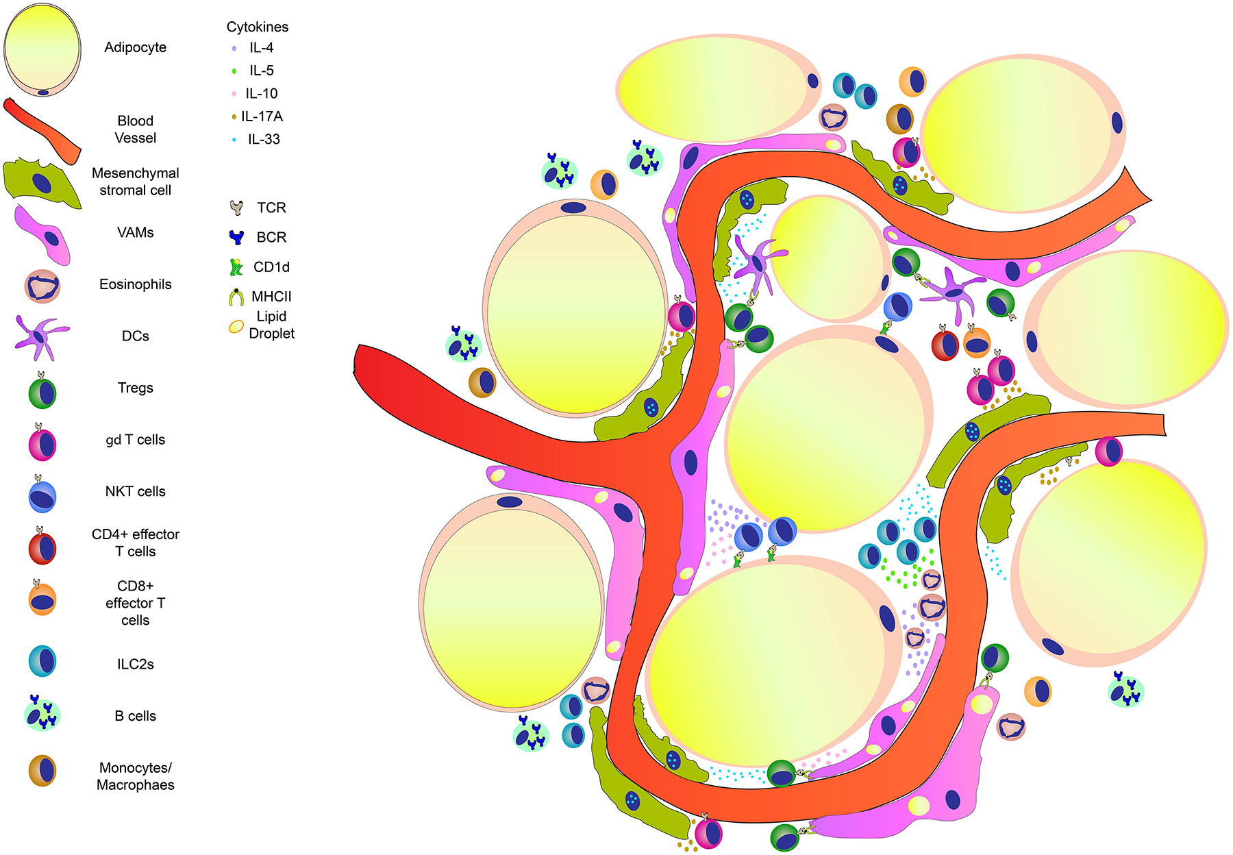
Figure 2. Major players of immune homeostasis in the lean eWAT.

The eWAT is an organ highly vascularized and replete of leukocytes. Vasculature associated adipose tissue macrophages (VAMs) are normally in intimate contact with the blood vessels. They display a rapid endocytic capacity for blood borne particles and also contain lipid droplets in their cytoplasm51. These cells are regulated by IL-4 and IL-10 produced by iNKT cells that are activated by CD1d expressing cells in the eWAT193. These cytokines contribute to an anti-inflammatory/ homeostatic environment shaping the activity of VAMs. γδ T cells positive for the transcription factor RORγT express high levels of IL-17A. This cytokine regulates the numbers of mesenchymal stromal cells (MSCs or Fibroblastic reticular cells)179. MSCs play an important role by producing IL-33, which is fundamental for the expansion of regulatory T cells (Tregs) within the adipose tissue. eWAT Tregs have a peculiar phenotype, displaying high expression of the receptor for IL-33157 and the transcription factor PPARγ154. Tregs can recognize antigens and be activated by VAMs or Dendritic cells (DCs). Upon activation Tregs release IL-10, which in turn contribute to the physiological eWAT environment. Another important cell population is the innate lymphoid cells type 2 (ILC2s)93. These cells are resident in a healthy eWAT producing high amounts of IL-5, which, in turn attracts and expands eosinophils. Production of IL-4 by eosinophils help to maintain the phenotype and activity of VAMs27. Other leukocytes reside in the eWAT (such as T cells, B cells, monocytes, macrophage and dendritic cells), however, their function in homeostasis remains to be investigated. TCR: T cell receptor; BCR: B cell receptor; MHCII- major histocompatibility complex 2.
