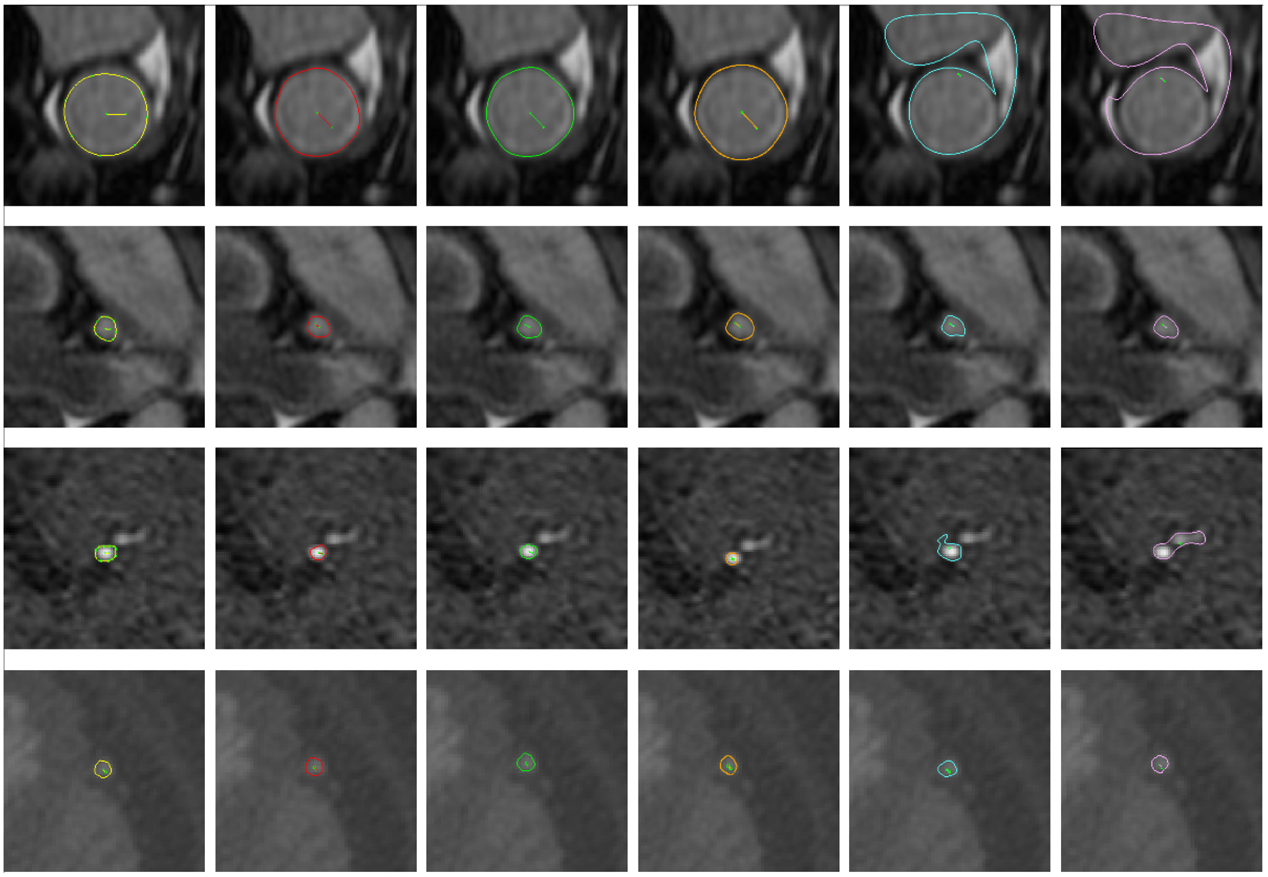
Fig. 6.

Selected example vessel surfaces detected by each method. From left to right: ground-truth, I2I-M, UNet-M, DeepLab-M, Threshold, and DLRS. From top to bottom: MR aorta, MR left coronary artery, MR posterior cerebral artery, and CT saphenous vein graft
