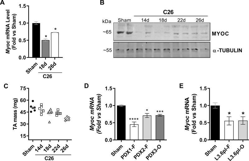
Figure 1. Myoc is transcriptionally downregulated in skeletal muscle in multiple experimental models of cancer cachexia.
A-C) Skeletal muscles harvested from C26 tumor-bearing mice were assessed for Myoc mRNA (A, TA muscles) and MYOC protein (B, gastrocnemius muscles) at various time points throughout tumor growth and the progression of muscle wasting (C, and Supplemental Fig. S1). Data are representative of n = 3–6 mice/group. D, E) The relative expression of Myoc mRNA was assessed in TA muscles in multiple experimental models of cancer cachexia, including in mice bearing human patient-derived xenograft (PDX) tumors implanted subcutaneously into the flank (PDX1-F, PDX2-F) or orthotopically into the pancreas (PDX-O) (D), and in mice inoculated with human L3.6pl tumor cells subcutaneously into the flank (L3.6pl-F) or orthotopically into the pancreas (L3.6pl-O) (E). Data are expressed as mean ± SEM, normalized to their respective sham group. Data are representative of n = 4–6 mice/group. For visualization purposes only, Sham groups for each cohort of PDX mice were combined, as were the Sham groups for the L3.6pl tumor-bearing groups. *P<0.05, **P<0.01, ***P<0.001, ****P<0.0001 vs Sham.

