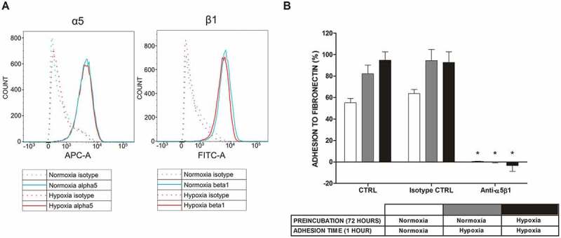
Figure 3.

Expression of α5β1 integrin in LAD2 mast cells at the protein level and mast cell adhesion to FN after blocking this receptor under normoxic and hypoxic conditions. (a) Expression of the α5β1 integrin receptor on the surface of LAD2 mast cells was assessed under conditions of 21% and 5/1% oxygen. A representative result of 21% (Normoxia) and 1% (Hypoxia) oxygen was selected. (b) Adhesion after 30 minutes of preincubation with 10 μg/mL anti-α5β1 antibody in 21% (Normoxia) and 1% (Hypoxia) oxygen. CTRL, control (untreated cells). Mean ± SEM. * p < 0.05, paired t-test.
