
Figure 5.
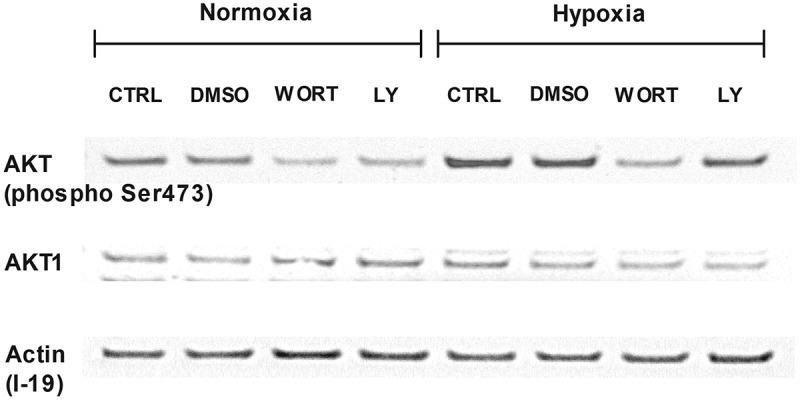
Involvement of the PI3 K/AKT signaling pathway in hypoxia. Western blot analysis of phosphorylated AKT and whole AKT1 and their expression in LAD2 mast cells. Cells were deprived of SCF for 72 hours. Then, protein isolates were collected after 30 minutes of preincubation with wortmannin (250 nM) or LY-294,002 (5 μM) and 1 hour in 21% (Normoxia) and 1% (Hypoxia) oxygen. CTRL, control (untreated cells); DMSO, cells preincubated with 0.05% DMSO; WORT, cells preincubated with 250 nM wortmannin; LY, cells preincubated with 5 μM LY-294,002.