Figure 3.

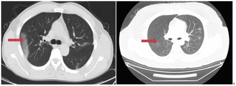
Typical changes in non-contrast enhanced chest CT scan. Multiple peripheral patchy ground-glass opacities with obscure boundary were seen in bilateral multiple lobes. Condensation shadow was observed on the lower right lobe.

Typical changes in non-contrast enhanced chest CT scan. Multiple peripheral patchy ground-glass opacities with obscure boundary were seen in bilateral multiple lobes. Condensation shadow was observed on the lower right lobe.