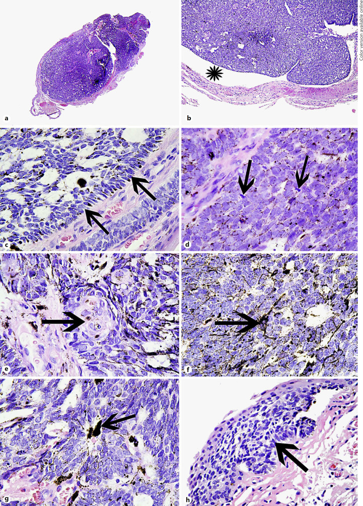
Fig. 2.
a Haematoxylin and eosin (HE)-stained section at scanning power of the BCC. b Artefactual processing cleft between the BCC and background conjunctival stroma. c HE. A peripheral palisade where the cells are lined up parallel to one another (arrows). d HE of the tumour cells showing mitotically active (arrows) tightly packed basaloid cells. Many tumour cells contain brown pigment in their minimal cytoplasm. e HE showing focal squamous differentiation (arrow). f HE. Arrow points to a dendritic pigmented melanocyte within the tumour. g HE. Arrow points to a melanophage in the tumour. h HE. The arrow points to a superficial pattern BCC connected to the under surface of the conjunctival epithelium. This corresponds to the smaller pigmented nodules flanking the main tumour (see Fig. 1a).

