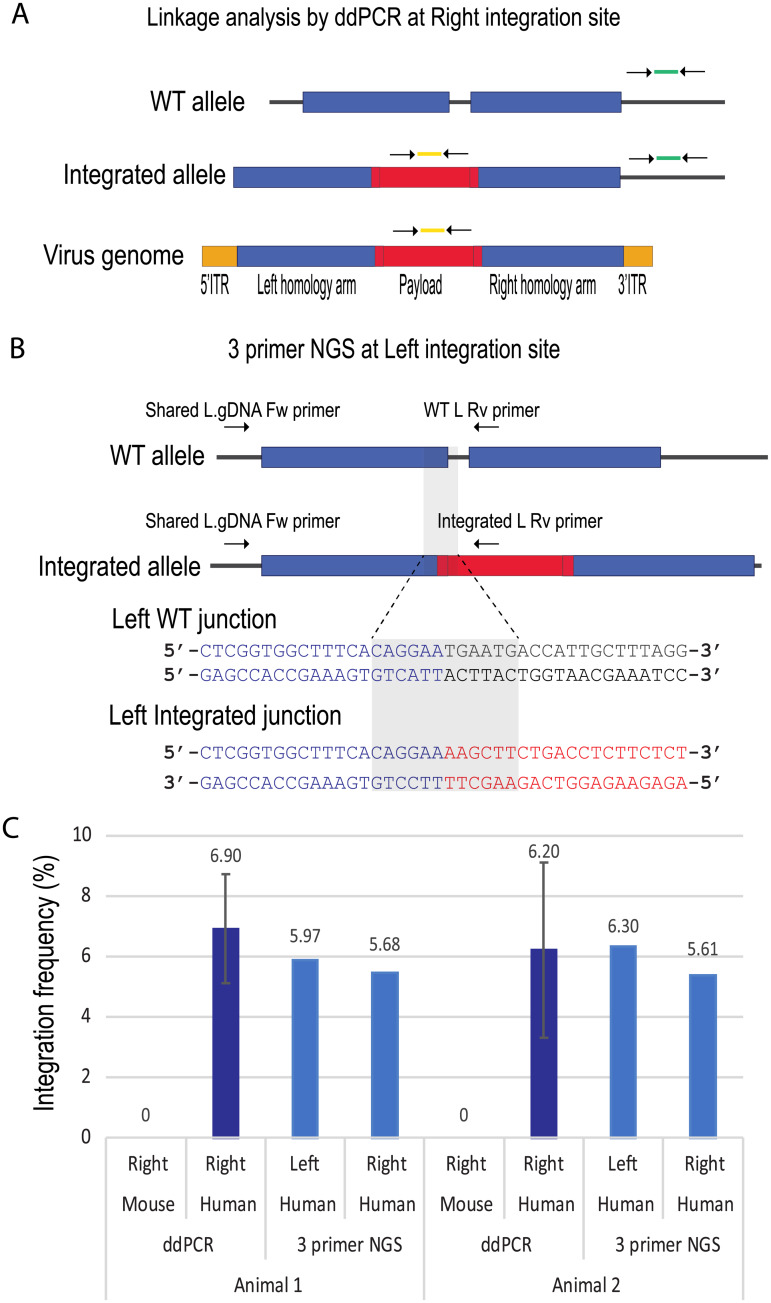
Fig 2. Integration frequency reaches 6% in human hepatocytes by two quantitation assays.
A. Schematic of linkage two-color ddPCR. Two primer-probe sets were used in this assay. FAM primer probe set (in yellow) detected coPAH payload, and HEX (in green) detected human genomic DNA outside of right homology arm. B. Schematic of 3 primer NGS assay at the left integration site. Three primers were used to amplify WT integration amplicon and integrated amplicon in a single PCR reaction. PCR products were sequenced with Illumina’s platform. The 12 base sequences unique for WT junction or for integrated junction are highlighted. The same assay was designed and performed at the right integration site. C. Linkage ddPCR and 3 primer NGS results of human genomic DNA or mouse genomic DNA. Note that a mouse specific HEX primer probe set was used for the mouse samples. In ddPCR, each sample was analyzed 3 times, and the error bar represents standard deviation. In 3 primer NGS, both left and right integration sites were queried.

