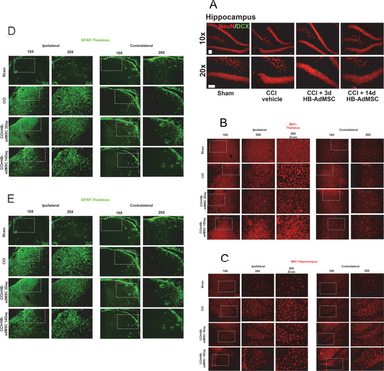
Fig 5. Representative localization of neuroinflammation and neurogenesis.
Thin sections from ipsilateral and contralateral hemispheres were immunostained at Day 32. Presented here are portions of the thalamus and hippocampus, specifically the subgranular zone (SGZ). A. Antibodies for NeuN and Doublecortin (DCX) were used to stain for neurogenesis, B, C. IBA-1 for microglial activation and D, E. GFAP for reactive astrocytes. Images are representative of sham, CCI + vehicle, CCI + HB-adMSCs 3d and CCI + HB-adMSCs 14d, at 20x magnification with a 10x inset showing a larger field. Scale bars indicate 200 μm.

