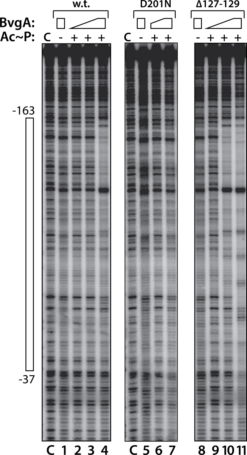
Fig 3. DNase footprinting of BvgA and derivatives on the pertussis toxin promoter.
DNase footprints were performed as described in Materials and Methods. BvgA proteins were wild-type (lanes 1–4), BvgAΔ201N (lanes 5–7), and BvgAΔ127–129 (lanes 8–11). Lanes designated “C” show the labeled fragment, digested with DNase I, in the absence of any added protein. Lanes 1, 5, and 8 show the digestion pattern obtained when BvgA was added but Ac~P was not. Acetyl phosphate was added to the reactions shown in the remaining lanes, as indicated. Concentrations of BvgA proteins used were 16 nM (lanes 2 and 9), 32 nM (lanes 3, 6, and 10) and 65 nM (lanes 1, 4, 5, 7, 8, and 11). The open bar to the left shows the maximal region protected in lane 11.

