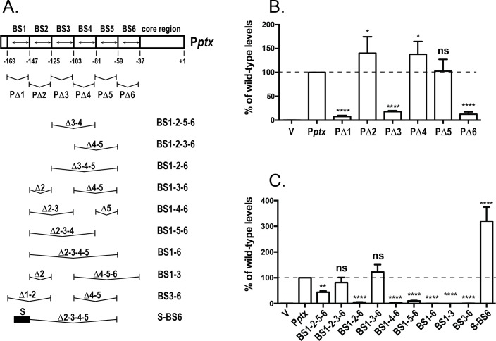
Fig 5. Contribution of Pptx BvgA-binding sites to Pptx function.
A. Diagram of BvgA-binding sites within Pptx and its derivatives. Boxes delineate the extents of 22-bp deletions removing each of the six binding sites, with the binding sites themselves within each box indicated by a double-headed arrow. Below this schematic are shown the different combinations of these 22 bp segments in different derivatives. The black bar in derivative S-BS6 indicates the presence of the primary binding site from Pfha (TAAGAAATTTCCTA). B & C. Luciferase activity of B. pertussis strain BP536 carrying ectopically integrated plasmids. Values for the empty pSS3967 control (V) and for promoter-lux fusion derivatives harboring the wild type (Pptx) and the deletion derivatives shown in panel A are presented. Strains were grown on BG agar at 37°C for 2 days and assayed as described in Materials and Methods. Values were normalized to wild-type Pptx and data from at least four assays were used in the calculation of means, standard deviations, as indicated by error bars, and statistical analysis by one-way ANOVA. Outcomes of the latter analysis are presented using the symbols: ns, P > 0.05; *, P ≤ 0.05; **, P ≤ 0.01; ***, P ≤ 0.001; ****, P ≤ 0.0001.

